
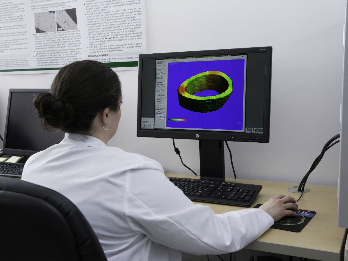
Micro-computed tomography, or microCT, is an X-ray-based imaging method that "sees" objects in three dimensions. The primary missions of the MicroCT and Histology Core are to provide state-of-the-art 3D ex vivo tissue imaging using microCT, and to provide histology services. Users can contract with the Core for microCT scanning and analysis, or can perform these activities themselves after required training and certification by Core personnel. We do not provide training for histology services. Please see our services brochure for further details.
Equipment Capabilities
Both our microCT scanners have software for data acquisition, online/offline reconstruction, 3D visualization and animation, as well as 2D and 3D trabecular bone histomorphometry. There is a database for tracking specimen-processing and archiving, with the capability to readily download histomorphometric data to spreadsheets or statistical packages.
- Desktop Cone-beam microCT Scanner Scanco model 45
- Nominal best resolution of 6 μm with a real resolution of 9 μm
- Field of view up to 37 mm and maximum scan length of 70 mm
- Nominal isotropic voxel size range is 1.25–100 µm
- Desktop Cone-beam microCT Scanner Scanco model 50
- Nominal best resolution 0.5 μm, with a real resolution of ~1 μm
- Field of view up to 48 mm; maximum scan length 120 mm
- Kubtec Parameter X-ray Cabinet with Digimus DEXA Software
- Planar X-ray imaging
- Dual Energy X-ray Absorptiometry (DEXA) measurement of bone mineral density (BMD) and body composition
- In vivo and ex vivo capabilities
- Maximum dimensions of specimen: 8” by 11” (203 mm by 279 mm)
- Magnification up to 3x available for improved detail
- Histology Preparation Equipment
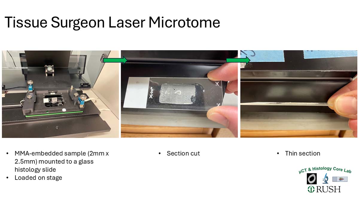
- Hard Tissue Histology preparation equipment includes methymethacrylate and epoxy embedding supplies, thin sectioning Tissue Surgeon Laser Microtome (LLS Rowiak) [Fig 1] [Fig 2], slab sectioning saws including Buehler Isomet 5000, Exakt 310 CP band saw, a Buehler Phoenix 4000 grinder/polisher
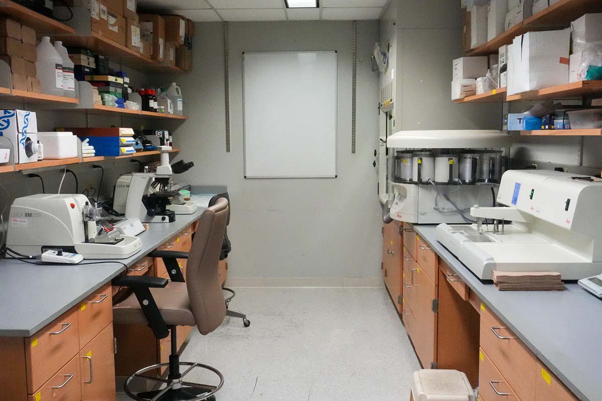
- Decalcified/soft Tissue Histology preparation equipment includes: Leica TP1020 automated paraffin processor, Arcadia paraffin embedder with cold plate, 2 Leica RM2255 microtomes
- Hard Tissue and Decalcified Soft Tissue section staining including H&E, Safranin-O, Masson's Trichrome, Basic Fucshin, Goldner's Trichrome, and others
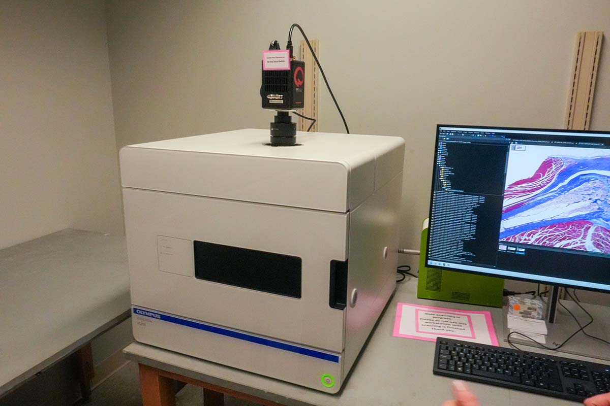
- Full section slide imaging using a Evident VS200 slide scanner [Fig 1] [Fig 2] [Fig 3]
Services Offered
MicroCT services
- Ex vivo X-ray micro-computed tomographic scanning of specimens
- 3D quantification of bone architecture, porosity and density
- 3D quantification of articular cartilage morphology and proteoglycan content
- 3D quantification of vascular networks
- 3D quantification of biomaterials
- Exportation of images (slices) in TIFF or DICOM format
- Creation of 3D renderings and animations of scanned objects
Kubtec services
- Ex vivo X-ray imaging, DEXA measurement of bone mineral density (BMD) and body composition
- In vivo X-ray imaging, DEXA measurement of bone mineral density (BMD) and body composition
Histology services
- Hard-tissue (undecalcifed) methylmethacrylate (MMA) processing and embedding
- Preparation of sections of MMA-embedded specimens, using the cut-and-grind method for histology and/or other specialized imaging techniques, such as backscatter scanning electron microscopy and Fourier transform infrared spectroscopy (FTIR) imaging. These specimens can contain metal implants.
- Preparation of thin sections of MMA-embedded specimens, using microtome-based methods
- Histologic staining of MMA-embedded sections (Basic Fuchsin)
- Decalcification, paraffin processing and embedding
- Preparation of sections using paraffin-embedded specimens
- Histologic staining of paraffin-embedded sections with hematoxylin and eosin stain (H&E), Toluidine Blue and others)
- Immunohistochemistry staining of paraffin-embedded sections
Expected Turnaround Times
Turnaround times are project-specific and dependent on current core instrument use. Please contact the core for an estimate.
Core Pricing, PO and Billing Information
Please review the following before requesting core services:
- Purchase Orders are required before services are rendered.
- Purchase Orders and Pre-Payment PO’s will not be valid for more than 365 days from receipt/invoice.
- A new PO must be submitted when current PO reaches $200 to avoid processing delays.
- Split PO requests must be submitted prior to services rendered. *A split PO request is defined as using multiple Purchase Orders to pay one invoice.*
- Accounts will be placed on hold when payments are not received within 120 days of the invoice date.
Please see this pricing document. Since many projects have custom needs, it is always best to contact core personnel to discuss pricing.
Past Clients Served / Current Collaborators
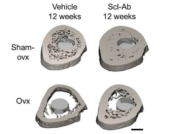
We have performed studies, or trained users, from Rush, the University of Illinois at Chicago, Northwestern University, the University of Chicago, Loyola University-Chicago, the University of Iowa, the University of Arizona, the University of Kentucky, and several companies. These projects have involved straightforward bone phenotyping (e.g., for genetic mouse models), studies of bone regeneration at various sites (long bones, cranial, ectopic), studies of bone-implant contact and peri-implant bone architecture, porosity of synthetic materials, vascular mapping (following contrast-agent perfusion), and articular cartilage imaging (with the use of a contrast agent).
Note: We ask clients to acknowledge use of this core in any published manuscripts.
Contact Us
Director for MicroCT and Hard Tissue Histology Cores
Meghan Moran, PhD
Phone: (312) 563-1257
Email: meghan_moran@rush.edu
Manager for MicroCT and Hard Tissue Histology Cores
Brittany M. Wilson, PhD
Phone: (312) 942-9809
Email: brittany_m_wilson@rush.edu