Our research mission is to advance translational research by paying attention to underlying mechanisms. The objective is to identify therapeutic targets and biomarkers which, after appropriate clinical investigation, will improve human health. Thus, projects run the spectrum from fundamental molecular biology and genetics to cell signaling to tissue, organ, and organismal level responses. Studies are conducted using cell culture, model organisms, and human subjects. There are four current areas of emphasis: cancer biology, musculoskeletal tissue injury and regeneration, movement disorders, and medical education research. ACB is the home department for the Joint Health NIH T32 training grant.
Principal investigators in Anatomy & Cell Biology integrate training opportunities into their research programs, including medical students, graduate students and post-doctoral fellows. Trainees are encouraged to perform research in cross-disciplinary areas and take advantage of opportunities in the clinical environment at Rush to develop basic research questions with a specific disease-oriented focus. The research typically involves collaborations not only with other researchers at Rush, but also with investigators elsewhere in the U.S. and abroad.
Anatomy & Cell Biology faculty direct three of the scientific instrumentation cores at Rush:
Cancer Biology
Projects involve osteosarcoma, breast cancer, lung cancer, lymphoma, ovarian cancer, uterine fibroids, leiomyosarcoma, cervical cancer, and bone metastases. Major pathways and approaches include p53, biomarkers, cancer cachexia, immunotherapy, autoimmunity, molecular-targeted ultrasound imaging agents, autophagy, growth factor signaling, cell metabolism, gene regulation, and prolylpeptidases.
- Laboratory of Jeffrey A. Borgia, PhD
- Laboratory of Lei Duan, MD
- Laboratory of Carl Maki, PhD
- Laboratory of Jitesh Pratap, PhD
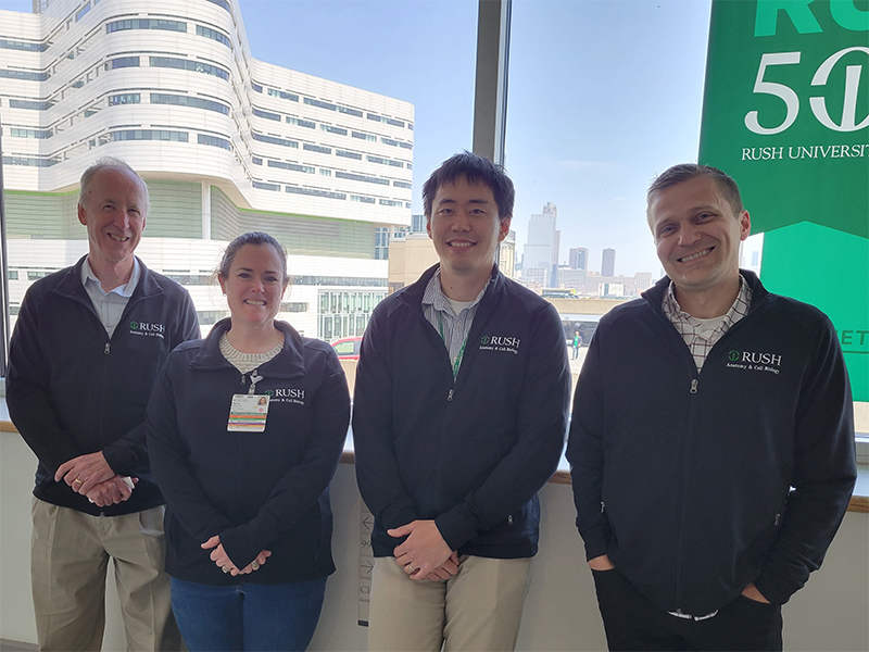
Musculoskeletal Tissue Injury and Regeneration: D. Rick Sumner, PhD, Meghan Moran, PhD, Frank Ko, PhD and Ryan Ross, PhD (left to right)
Musculoskeletal Tissue Injury and Regeneration
Projects include implant fixation, joint replacement, osteoarthritis, osteoporosis, large bone defects, bone and cancer cell interaction, osteomalacia, and Alzheimer’s Disease. Major pathways and approaches include bone regeneration, bone remodeling, biomarkers, gene and protein expression, Wnt signaling, stem cell biology, skeletal mineralization, and mineral metabolism.
- Laboratory of Frank Ko, PhD
- Laboratory of Meghan Moran, PhD
- Laboratory of Ryan Ross, PhD
- Laboratory of D. Rick Sumner, PhD
- Laboratory of Amarjit S. Virdi, PhD
- Laboratory of Brittany M. Wilson, PhD
Movement Disorders
Projects involve Fragile X tremor/ataxia syndrome (FXTAS), Parkinson’s Disease, Huntington’s Disease, and Niemannn-Pick Type C. Major pathways and approaches include early detection of disease onset and treatment efficacy through study of balance, gait and functional mobility.
Medical Education Research
Projects tend to be focused in the area of ‘educational measurement and evaluation’. This entails a range of topics including test/instrument development and validation, program evaluation, and quantitative analyses of educational and psychological data directly pertinent to medical and anatomy education.