
Manno-Figueroa Research Group
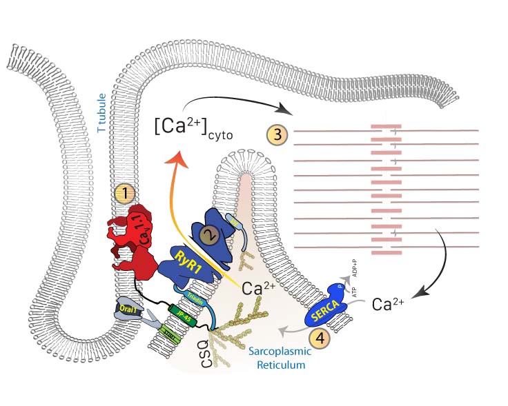
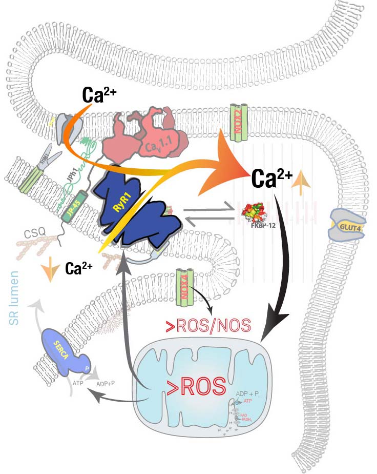
Our laboratory (https://myolab.weebly.com) investigates how the dynamic movement of calcium between intracellular compartments orchestrates the transition from muscle rest to contraction in skeletal muscle (Figure 1). We focus on the proteins that regulate calcium release and reuptake, how defects in these molecular systems disrupt calcium homeostasis, and the mechanisms by which these disturbances drive the development of myopathies (Figure 2).
Our research spans a broad spectrum of muscle disorders, including malignant hyperthermia (MH), central core disease (CCD), Duchenne muscular dystrophy (DMD), sarcopenia, and chemically-induced myopathies, as well as hyperglycemia and diabetes. By uncovering the fundamental mechanisms that underlie these conditions, we aim to identify novel therapeutic strategies that preserve muscle function and ameliorate muscle disease.
Our Work
Our research group has made significant strides in elucidating the mechanisms of calcium dysregulation in skeletal muscle, particularly through our work on "couplonopathies" --diseases caused by defects in the multi-protein unit responsible for intracellular calcium release. We have identified "chemical couplonopathy" as a key factor in doxorubicin-induced skeletal myopathy (DISM), in which chemotherapy triggers a destructive cycle of calcium leak and oxidative stress. To address this, we have identified novel use-dependent RyR1 inhibitors, such as MP-001 and R-carvedilol, that specifically target abnormal channel activity to restore muscle force and fatigue resistance.
In the context of inherited myopathies, such as CCD, MH, and DMD, we are studying the extent of RyR1 receptor dysfunction (calcium leak) in each scenario using patient-derived cells and animal models. Our long-term goal is to identify disease-specific RyR1 inhibitors to improve the quality of life for affected subjects in each case. We are also using the same approach to try to improve resistance to fatigue during aging. Our preliminary findings are promising and suggest that these approaches may help improve movement quality and muscle strength in animals.
Beyond muscle performance, we have pioneered research linking muscle calcium stress to metabolic disorders, including type 2 diabetes. Our studies demonstrate that chronic elevations in resting cytosolic calcium impair glucose storage and promote hyperglycemia. We discovered that this calcium stress can cleave proteins like junctophilin-1, triggering gene regulatory programs that attempt to correct glucose dysregulation. These findings highlight the essential role of maintaining calcium homeostasis in skeletal muscle in preventing insulin resistance, particularly in aging and various muscle diseases.
Technology and Methods
Our lab uses electrophysiological techniques, imaging of live specimens, and optical monitoring of function, combined with ancillary molecular biology and protein chemistry.
In addition, we have developed novel techniques and approaches:
- A biorepository of patient-derived muscle cells and tissues
- Interpretation of experimental data using quantitative modeling and simulation
- A novel method to simultaneously measure calcium & force in the whole muscle
- A biosensor for imaging Ca concentration in the sarcoplasmic reticulum
- A novel method for determining murine skeletal muscle fiber type using autofluorescence lifetimes (Fluorescence Lifetime Imaging Microscopy)
- 3D-printed flow chambers for combining multiple operations in single- or multi-cell preparations, or cell cultures, including fast laminar perfusion, electrical stimulation, patch clamp, and thermal control (Patent PCT/US2016/026097)
Publications
Last 5 years
- Figueroa LC, Tammineni ER, Marco-Moreno P, Vallejo-Illaramendi A, López de Munain A, Sagartzazu-Aizpurua M, Fill M, Manno C. Disrupting the Feed-Forward Cycle of RyR1 Ca Leak and Oxidative Stress Mitigates Doxorubicin-Induced Skeletal Myopathy. Am J Physiol Cell Physiol. 2026 May 19; PubMed PMID: 42154967.
- Tammineni ER, Figueroa L, Manno C. Calpain Mediated Proteolysis of Junctophilin-1 Produces an Aggregation Prone C-Terminal Fragment in Skeletal Muscle. Res Sq. 2026 Feb 3; PubMed PMID: 41674813.
- Tammineni ER, Figueroa L, Fill M, Riazi S, Manno C. Age-Dependent Effects of Muscle Resting Calcium on Fasting Blood Glucose: Implications for Prediabetes Risk. Endocrinol Diabetes Metab. 2025 May;8(3): e70052; PubMed PMID: 40329491.
- Tammineni ER, Manno C, Oza G, Figueroa L. Skeletal muscle disorders as risk factors for type 2 diabetes. Mol Cell Endocrinol. 2025 Apr 1; PubMed PMID: 39848431.
- Ríos E, Samsó M, Figueroa LC, Manno C, Tammineni ER, Rios Giordano L, Riazi S. Artificial intelligence approaches to the volumetric quantification of glycogen granules in EM images of human tissue. J Gen Physiol. 2024 Sep 2;156(9); PubMed PMID: 38980209.
- Figueroa L, Kraeva N, Manno C, Ibarra-Moreno CA, Tammineni ER, Riazi S, Rios E. Distinct pathophysiological characteristics in developing muscle from patients susceptible to malignant hyperthermia. Br J Anaesth. 2023 Jul;131(1):47-55; PubMed PMID: 36792386.
- Tammineni ER, Figueroa L, Manno C, Varma D, Kraeva N, Ibarra CA, Klip A, Riazi S, Rios E. Muscle calcium stress cleaves junctophilin1, unleashing a gene regulatory program predicted to correct glucose dysregulation. Elife. 2023 Feb 1;12; PubMed PMID: 36724092.
- Manno C, Tammineni E, Figueroa L, Oropeza-Almazán Y, Rios E. A novel method for determining murine skeletal muscle fiber type using autofluorescence lifetimes. J Gen Physiol. 2022 Sep 5;154(9); PubMed PMID: 35796671.
- Manno C, Tammineni E, Figueroa L, Marty I, Ríos E. Quantification of the calcium signaling deficit in muscles devoid of triadin. PLoS One. 2022;17(2):e0264146; PubMed PMID: 35213584.
Complete list of Dr. Figueroa’s published works:
https://www.ncbi.nlm.nih.gov/myncbi/lourdes%20%20carolina.figueroa.1/bibliography/public/
Complete list of Dr. Tammineni’s published works:
https://scholar.google.com.mx/citations?user=4JRJ7TcAAAAJ&hl=en
Complete list of Dr. Manno’s published works:
https://www.ncbi.nlm.nih.gov/myncbi/carlo.manno.1/bibliography/public/
Funding
Recently completed
- Cohn Fellowship, Rush Research Mentoring Program, Rush University (Total cost: $25,000.00)
* Title of Project : Use-dependent inhibition of RyR1 channels, a promising therapy for age-related muscle wasting.
* Period: July 2025-June 2026 - Rush Translational Sciences Consortium (RTSC), Rush University (Total cost: $100,000.00)
* Title of Project: Deciphering the Molecular Mechanisms Underlying Calcium-Dependent Disruption of Skeletal Muscle Excitation-Contraction Coupling During Cancer Chemotherapy and Exploring Strategies for Symptom Alleviation
* Period: Feb 2025- January 2026
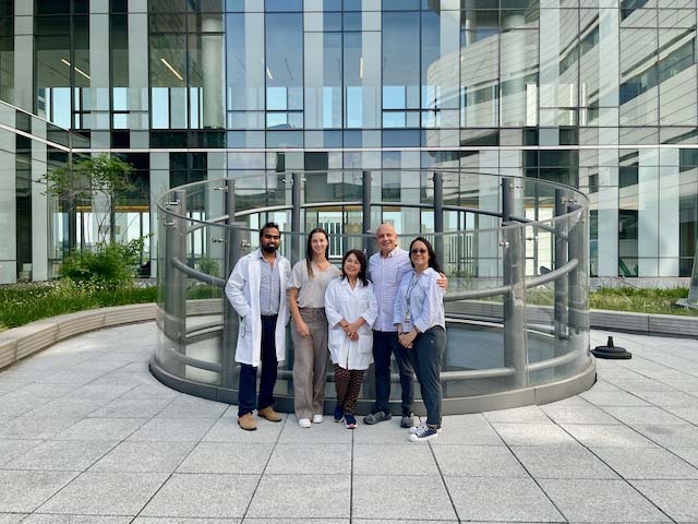
Our Team
Lourdes Carolina Figueroa, Ph.D. (Assistant Professor)
Eshwar Tammineni, Ph.D. (Instructor)
Carlo Manno, Ph.D. (Assistant Professor)
Contact Us
Carolina Figueroa, PhD
Jelke Building, Room 1267
Phone: (312) 942-4458
Email: lourdes_figueroa@rush.edu
Eshwar Tammineni, PhD
Jelke Building, Room 1279
Phone: (312) 942-8044
Email: eshwarreddy_tammineni@rush.edu
Carlo Manno, PhD
Jelke Building, Room 1271
Phone: (312) 942-4464
Email: carlo_manno@rush.edu