New devices and technology are advancing cardiac diagnosis and treatment
A change of heart can be a romantic disaster, especially on Valentine’s Day, but ongoing changes in heart care are working miracles for patients with cardiac conditions. New and smaller medical devices, advances in minimally invasive surgery and more powerful imaging technology are extending and enhancing lives, bringing innovative treatments to wider groups of patients, and diagnosing conditions more accurately and quickly.
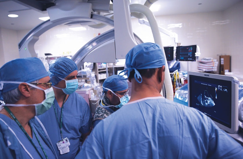
The Rush System is helping lead these improvements in heart care by conducting clinical trials of new treatments and being the first area health care provider to offer new options. Here, doctors from each of Rush’s three hospitals discuss some of these innovations and how they’re benefiting patients.
Closing a tiny gap that causes big problems
The heart is a pump, and like any pump it has valves. One of them, the mitral valve, is located on the upper and lower chambers on the heart’s left side, the left atrium and the left ventricle, and regulates the flow of oxygenated blood.
“Because we’re living longer, we’re seeing more age-related disorders, and one of the most common age-related disorders is a disorder of the mitral valve,” says Clifford Kavinsky, MD, PhD, an interventional cardiologist and director of the Rush Center for Adult Structural Heart Disease.
The mitral valve can leak due to a variety of problems that cause the heart to become enlarged, so the valve doesn’t close properly, allowing blood to flow backwards from the atrium into the ventricle. Known as mitral regurgitation, this condition about one in 10 adults more than 75 years old and can cause shortness of breath, fatigue and lightheadedness and can be life-threatening.
Doctors can perform open-heart surgery to repair or replace the valve, but elderly patients and others at high risk may be too frail to undergo the surgery. Kavinsky offers an alternative treatment, MitraClip, a device inserted through an incision in the groin and threaded through the blood vessels to the heart, where it’s inserted between the leaflets of the mitral valve.
“We draw the leaflets together with the clip, and it leaks less,” Kavinsky explains.
In 2013, the FDA approved MitraClip for patients born with an abnormal, leaky mitral valve who were too sick for surgery. Kavinsky also is able to offer MitraClip for heart failure patients with mitral regurgitation because he’s been part of a national study of the treatment, the COAPT trial.
“We’re doing it for three or four patients a month,” he says. “You significantly reduce the leakage, patients no longer have symptoms of heart failure, and it’s far safer than cardiac surgery.”
Close to the heart
Doctors use pacemakers to help control irregular heartbeats, especially abnormal heart rates, by sending electrical pulses to the heart. Most pacemakers are implanted underneath the left clavicle (collarbone) and attached to the heart using wires called leads, which are threaded through a vein to reach the heart. While the pacemaker helps maintain normal heart beat in people with slow heart rates, the wires limit motion and are a risk for infection.
“The wires go through your pectoral muscle underneath your clavicle. As that muscle gets stretched, over time it can literally ratchet out the lead,” explains Edward Lipman, MD, an electrophysiologist at Rush Copley Medical Center.
The first leadless pacemaker, Micra, which is about the size of a vitamin capsule, is implanted directly in the heart, avoiding the complications of much larger, conventional pacemakers. “You can be active. You don’t have to worry about dislodging the leads,” says Lipman, who in March of 2018 became the first doctor in the Fox Valley area to implant the Micra pacemaker.
Micra is what’s known as a single-chamber pacemaker and is used in a ventricle, one of the heart’s bottom chambers. “It’s good for people fainting from sudden pauses in heartbeat, or constant atrial fibrillation, where they go 10 seconds without a beat,” Lipman says.
The FDA approved Micra in April 2016, but doctors who weren’t involved in clinical trials of the device only could begin receiving training to use it until late 2017. “I did the second or third training session,” Lipman says.
Unlike conventional pacemakers, which are inserted through an incision in the chest, Micra pacemakers are implanted using a non-invasive procedure. Doctors attach it to a catheter, which they pass through an incision in the upper thigh and up to the heart to put the pacemaker in place before withdrawing the catheter.
A high-tech look into the heart
When doctors think a patient may have heart disease due to their symptoms or risk factors, they may order a stress test to further assess the patient’s condition. Many patients will get an exercise stress test — walking or running on a treadmill while sensors monitor their heartbeat — but patients with physical limitations instead may receive a test using nuclear imaging.
Rush recently acquired the technology to perform these tests using PET perfusion, an advanced nuclear imaging technique that provides greater diagnostic accuracy than traditional nuclear imaging using SPECT technology. (PET stands for positron emission tomography and SPECT is short for single-photon emission computed tomography.)
“Currently, we look at relative blood flow which has limitations. With PET, we can actually measure the blood flow going into the tissue,” says Rupa Sanghani, MD, director of nuclear cardiology at Rush.
PET scans also take far less time than SPECT imaging — 35 to 45 minutes versus three hours — and uses less radiation. “It’s more efficient for everyone involved, easier for the patient, less radiation exposure for the patient, and you get better quality results,” Sanghani says.
PET imaging has been in use in cancer diagnosis for about 10 years, but it’s rarely used in cardiology due to the expense of the equipment and materials involved and the expertise needed to use the technology. Rush will have the only stand-alone cardiac PET imaging program in the Chicago area. The program will begin imaging in late February, with wider availability in the spring.
“We’re very excited to offer cardiac PET. It’s state-of-the-art technology and will provide robust clinical information to Rush’s patients and patients being referred to us,” Sanghani says.
Life-saving new technology at Rush Oak Park Hospital
Cardiac care technology also continues to advance at Rush Oak Park Hospital, where construction soon will be completed on a new dual energy CT scanner that will serve the hospital’s Emergency Department.
Not only will the new CT scanner provide better resolution of images and less radiation exposure to patients, but it also will include enhanced cardiac capabilities called fractional flow reserve computed tomography, or FFR-CT. This technology allows doctors to determine whether blockage areas in coronary arteries are depriving the heart of blood flow that can lead to a heart attack or sudden death.
"This technology can be a life-saver for a patient who comes to our emergency room with possible heart issues,” said Donald Tanis, MD, a cardiologist at Rush Oak Park Hospital. “With this technology we can design treatment plans without performing invasive procedures.”
Annabelle Volgman, MD, a cardiologist at Rush and medical director of the Rush Heart Center for Women, said this technology is vital for women experiencing heart issues: “FFR-CT is especially helpful for women since they have heart disease that is difficult to diagnose without this advanced technology.”
According to LuAnne Smith, director of medical imaging and cardiology at Rush Oak Park Hospital, the CT scanner’s enhanced cardiac imaging capability is expected to be up and running in April. “Rush Oak Park Hospital will be the only hospital in Oak Park with this level of cardiac imaging technology,” she says.
Expanding the reach of a recent advance
“Transcatheter heart valves have been FDA approved for less than a decade, so we don’t know how long they’ll last,” says Antone Tatooles, MD, chief of cardiac surgery at Rush. Made from the lining of cow hearts, they’re used to replace a narrowed aortic valve — which controls the flow of blood from the heart — and are guided into place on a catheter passed through the skin like an IV and threaded to the heart.
“We don’t have to open their chest. The valve can be delivered through the groin, through the arm or through the bottom of the heart,” Tatooles says. This minimally invasive approach, called transcatheter aortic valve replacement,. allowed doctors to replace aortic valves in patients who were poor risks for surgery, such as older adults
“As the valves and the delivery systems get better, we’re finding different patient populations. The valves that we're using today at Rush are valves that the FDA already approved, but the patient population who we're using them on is different. We’re trying to better define who are the most appropriate patient for this therapy.”
Tatooles has been part of studies examining whether patients with low risk of surgical complications, particularly younger adults are appropriate for transcatheter valves rather than surgical valves. “Time will tell, because we’re still evaluating the durability of these valves,” he says.